
Neutron Imaging and Tomography Facility
For more information, please contact Boris Khaykovich.
The MITR utilizes a 4" thermal neutron beam port for neutron tomography and radiography. A range of apertures is used to achieve the spatial resolution necessary for the sample being imaged.
The facility uses a standard scintillator-based detector equipped with a commercial scintillator (Lexel Imaging) measuring 3.75"x 3.75", a lens, and a charge-coupled device (CCD). A sample can be installed on the rotation stage available for tomography and a spatial resolution of 30 µm can be achieved.
Figure 1 (below) shows the imaging facility with a sample (stack of coin cell batteries) on the rotation stage. The scintillator, a 20 µm thick deposit of P43 GadOx on a 0.020" thick aluminum substrate (1100 series), is inside a light-tight box behind an aluminum front plate. The box also contains a mirror for deflecting the light away from the neutron beam.
The light-tight bellows between the scintillator box and the CCD contains a Nikon photographic lens. The CCD is a ZWO ASI1600 Pro monochrome cooled CMOS astro camera with a 3.8 µm pixel size. The Nikon lens achieves a range from pixel size of 5.24 µm with field-of-view (FOV) of 24x18 mm2 to a pixel size of 17.56 µm with FOV of 81x51 mm2 (more detector parameters).
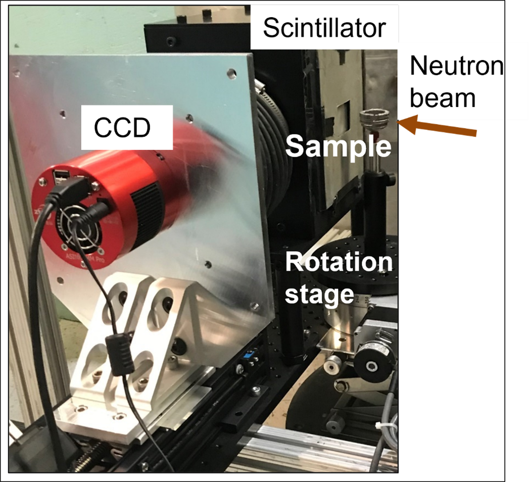
Figure 2 (below) shows an example of tomographic imaging of a 3-D printed stainless-steel object. Other previous projects include:
- imaging of irradiated hydride fuel rodlets
- in-situ radiography of electrolyzer cells
- dynamical imaging of water boiling
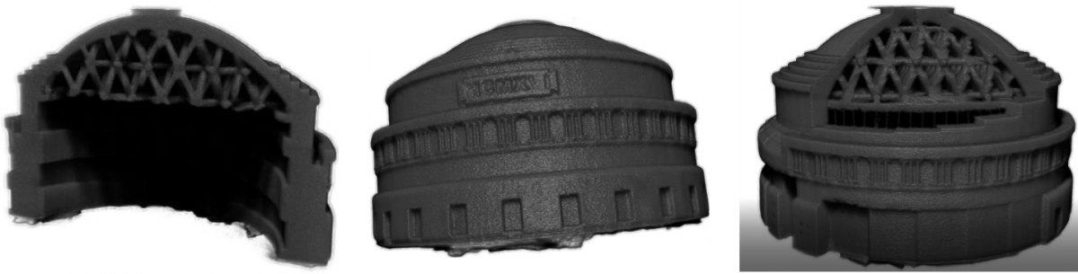
Figure 2: Tomographic projections of a 3-D printed model of the MIT Great Dome structure. The model is made of stainless steel and is 1" in diameter
Figure 3 (below) shows tomographic images of three different solid-state electrolyte batteries. Such batteries are much safer than traditional Li-based batteries that use flammable liquid electrolyte. Neutron tomography improves the ability to see lithium plating during battery cycling, and so to optimize the materials composition (Li in ceramic batteries is difficult to image with X-rays due to high-Z ceramic materials masking the signal from low-Z Li.)
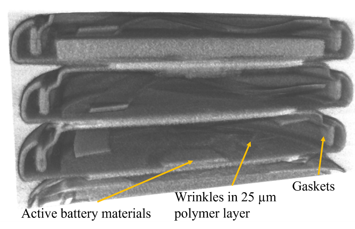 Figure 3: Tomographic images of solid ceramic electrolyte batteries with their parts
Figure 4 (below) shows 3D printed aluminum oxide particles in polymer binder. Parts are heated to sinter particles and burn out the binder. Each small cylinder was subjected to a different temperature (as formed at top) to determine onset of binder burn-off. Changing contrast follows changes in the quantities of the remaining polymer binder.
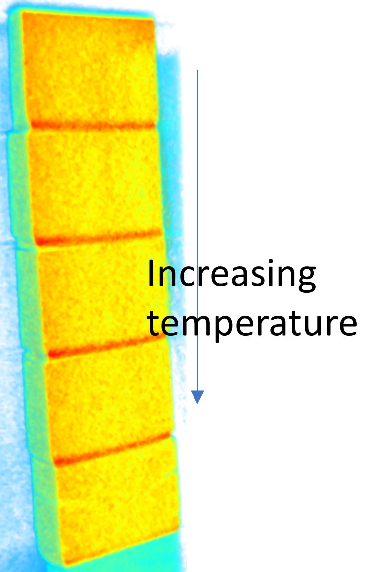
Figure 4: Tomographic image of a stack of 3D printed aluminum oxide composites sintered at different temperatures
Detector Parameters
- Camera: ZWO ASI 1600MM Pro monochrome cooled CMOS astro camera. Pixel size is 3.8 microns on the chip.
- Lens: Nikon Nikkor f/1.4 85mm lens with extension tubes. Currently installed is a PK13 extension tube. The installed lens configuration gives a pixel size of 8.74 micron with a field of view of 40 mm x 30 mm. It is possible to achieve a range from a pixel size of 5.24 micron with FOV of 24 mm x 18 mm to a pixel size of 17.56 micron with FOV of 81 mm x 51 mm by changing the extension tube configuration. With a Nikon 50mm lens you can see the entire scintillator with a FOV of 139 mm x 105 mm.
- Scintillator: The phosphor screen was manufactured by Lexel Imaging in Lexington Kentucky. It has an active area of 3.75" x 3.75". The coating is a 20 micron thick deposit of P43 GadOx on a 0.020" thick 1100 series aluminum substrate. The resolution of the scintillator is most likely in the 15-20 micron range based on our older scintillators.
- Resolution: The detector pixel size is currently set at 8.74 micron giving a best case resolution of 17.5 micron, which falls around the limitations of the scintillator. Given the L/D of 517 and a sample to detector spacing of 25 mm, the true resolution is likely closer to 30-40 microns. In theory, if a smaller sample diameter is used so that the sample-to-detector distance can be reduced to 10 mm, the resolution would improve to 20 microns.
For more information, please contact Boris Khaykovich.